 |
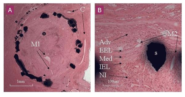
| Figure 5: (a) Section from a pig coronary at 28 days (hematoxylin and eosin). The neointima is mature, and completely occludes the lumen. ‘Large’ microvessels (100-200μm diameter) are visible in the centre of the occlusive neointima (M1) and similar sized collateral vessels are seen in the adventitia around the stented segment (C). (b) High power view of the neointima around a stent strut from another animal at 28 dyas. The neointima (NI), internal elastic lamina (IEL), media (Med), external elastic lamina (EEL) and adventitia (Adv) are labelled. ‘Small’ microvessels (10-20μm diameter) are visible in the neointima around the strut. The neointima of the CTO appears to be composed of the typical vascular smooth muscle cells and matrix. |