 |
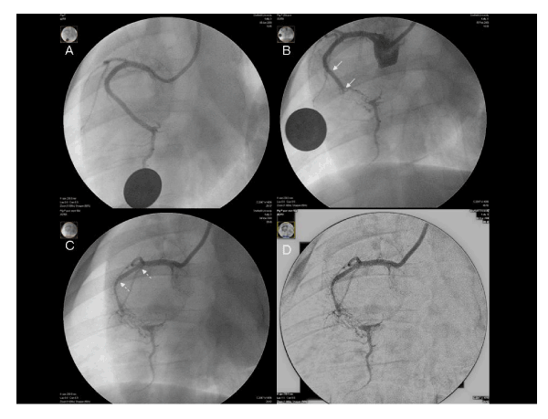
| Figure 6: Angiographic images from a control group animal. A Initial RAO 30° view of RCA on day 0 prior to implantation of copper stent. B Same view at day 28 demonstrating mid vessel CTO at distal edge of copper stent (solid arrows) with antegrade bridging collaterals. C Day 56 following implantation of study (control, PEP polymer only) stent (dashed arrows). D Digital subtraction image of C used to highlight microvessels for image analysis. |