|
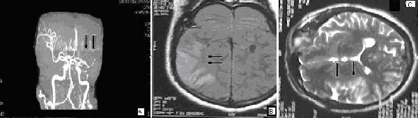
| Figure 3: A. Cranial MRI angiography shows that peripheral arteries of left MCA are not clearly visualized compared to right ones and peripheral flow of the artery must be diminished probably due to an occlusive lesion in the proximal portion. B, C. Cerebral MRI shows a wide high intensity area in the right fronto-temporoparietal region (Coronal FLAIR and axial T2W sequences). |