|
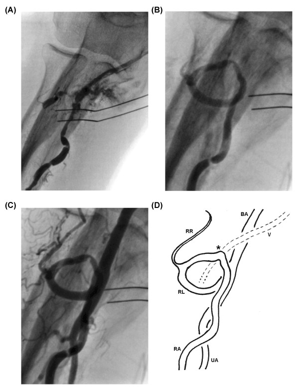
| Figure 2: (A) Angiogram showing perforation of the radial artery with contrast extravasation and crossing into an adjacent vein. (B) and (C) Retrograde angiograms showing a complete radial artery loop. (D) Schematic vascular anatomy of the antecubital region; BA, brachial artery; RA, radial artery; UA, ulnar artery; RL, radial loop; RR, recurrent radial artery; V, side vein; *, point of perforation at the radial loop vertex and crossing into adjacent vein. |