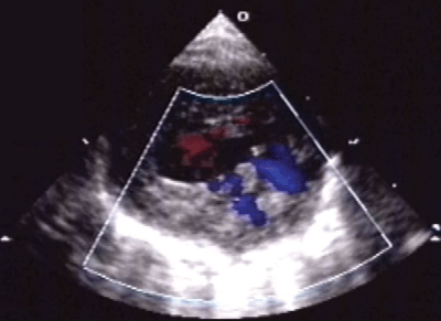
Figure 1:
PSAX view showing cross section of LV and RV with prominent trabeculations, deep intertrabecular recesses with colour flow (seen as blue) continuous with the ventricular cavity.