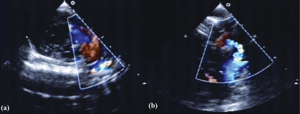
Figure 3:
(a)
Modified PSAX view showing PDA (asterisk) shunting right to left.
(b)
Suprasternal long axis view showing hypoplastic aortic arch with area of turbulent flow followed by dilated segment in descending aorta.