 |
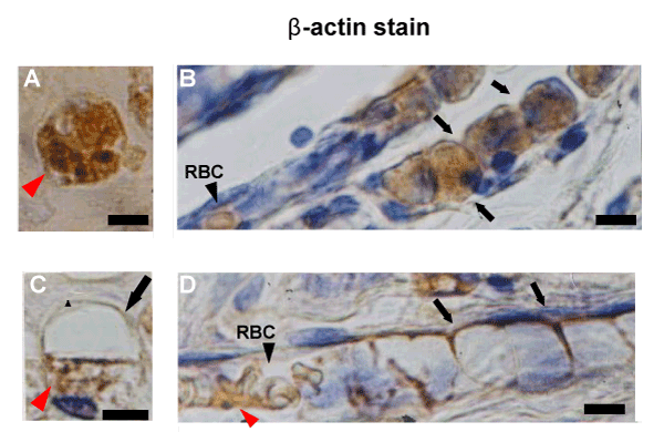
| Figure 4: β-MNCs adherent to each other in tandem. A: Transected view of a β-MNC. The cell was filled with β-actin-positive granules (red arrowhead). B: Longitudinal view of β-MNCs filled with β-actin-positive granules and adherent to each other in tandem (arrows), indicating an immature capillary tube. Black arrowhead: A red blood cell (RBC) drained into a proximal segment of the capillary tube where intercellular junctions were lost. Two weeks after infarction production. C: Transected view of a β-MNC with a cavity (arrow) and residual granular cytoplasm (red arrowhead). D: Longitudinal view of a capillary tube in which granular cytoplasm were lost almost completely. Arrows: persistent intercellular junctions. RBCs appeared in a proximal portion of the tube where the intercellular junctions had disappeared (black arrowhead). Red arrowhead: residual β-actin-positive granular cytoplasm. Four weeks after infarction production. |