|
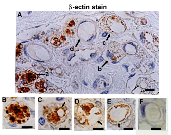
| Figure 5: Various stages of cavity formation in β-MNCs. A: β-actin stain 4 weeks after myocardial infarction. a: A β-MNC filled with β–actin-positive granules. b: Formation of a small cavity. c: Loss of granular cytoplasm and formation of a large cavity. d: Complete loss of granular cytoplasm, with a smoothed luminal surface. Degranulation, cavity formation and consequent maturation were considered to occur in the order of B to F. Four weeks after infarction production. |