|
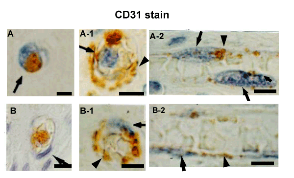
| Figure 6: Two different configurations of nuclei of CD31-positive MNCs that formed capillary tube. A: A MNC containing a round nucleus and CD31-positive granular cytoplasm (arrow). A-1: An EC forming a tube. CD31 was present in the cell membrane (arrowhead). The cell possesses a round nucleus (arrow). The tube contained red blood cells. A-2: Longitudinal view of a capillary tube. The ECs possess round nuclei (arrows) and CD31 in the cell membrane. B: A MNC containing a flat nucleus (arrow) and CD31-positive granular cytoplasm (arrowhead). B-1: An EC with a flat nucleus (arrow) forming a tube. CD31 was present mainly in the cell membrane (arrowhead). A-2: Longitudinal view of a capillary tube. The ECs have flat nuclei (arrow) and CD31 in the cell membrane (arrowhead). |