|
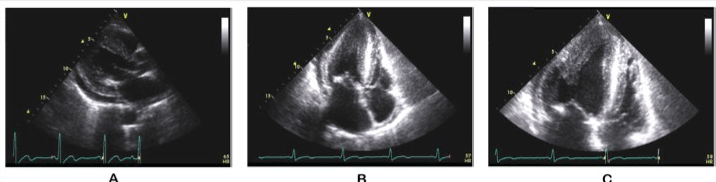
| Figure 2: Transthoracic echocardiogram. A. Parasternal long axis view showing thickened septum and posterior wall of the left ventricle. B & C. Four chamber apical view showing thickened left ventricular walls with asymmetric involvement at the apex. Pericardial effusion and left atrial enlargement is also seen. |