|
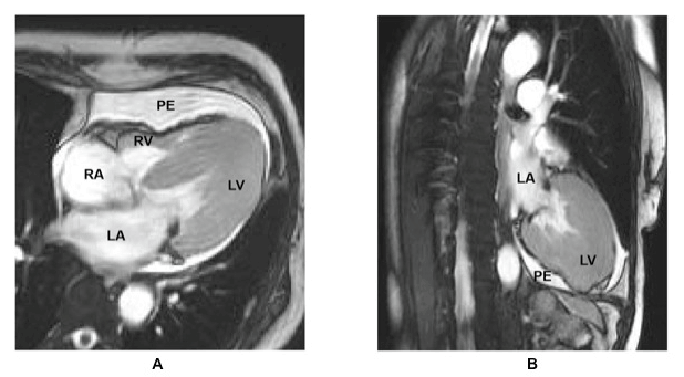
| Figure 3: Cardiac MRI. A. Four chamber axial and B. two-chamber views using steady state free precession sequence are shown. There is asymmetric hypertrophy of the LV that is most prominent at the apex with a spade-like configuration of the LV cavity consistent with the apical variant of Hypertrophic Cardiomyopathy (HCM). There is also a moderate circumferential pericardial effusion. The Left Atrium (LA), Left Ventricle (LV), Pericardial Effusion (PE), Right Atrium (RA) and Right Ventricle (RV) are labeled. |