 |
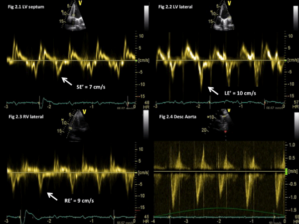
| Figure 2: Tissue Doppler imaging of the septum (2.1), lateral LV (2.2), lateral RV (2.3), and respiratory variations on the descending aorta (2.4). Note the preserved diastolic myocardial velocities, although with a higher value for the basal lateral LV than the septal LV. |