|
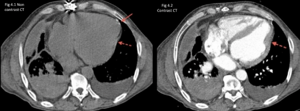
| Figure 4: Computed tomography scans of the thorax (with and without contrast). Dashed arrow: localized thickening in the mid infero-lateral LV wall (6.4 mm). Solid arrow: pericardial calcification. There is also right-sided pleural thickening with bilateral pleural effusion (organized in the right pleural cavity). |