|
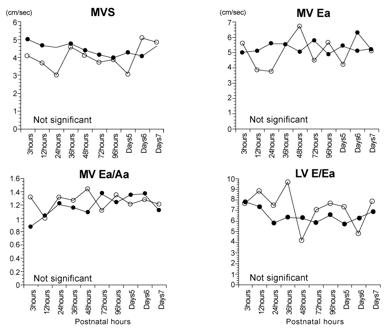
| Figure 2: Longitudinal comparisons of tissue Doppler velocities in the lateral portion of mitral valve (MV) anulus in the early neonatal period between infants in the recipient group (open circle) and infants in the control group (closed circle). Values are median. |