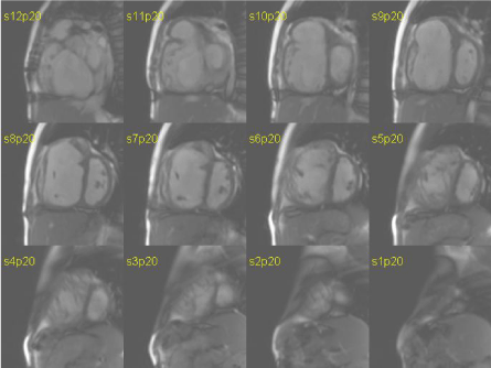
Figure 1:
True fast imaging with steady-state precession sequence in short axis orientation at medium level during diastole (right) and sistole (left) in a patient with pressure and volume right ventricle overload.