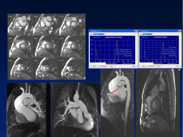
Figure 4: Up-left: True fast imaging with steady-state precession sequence
of short axis in a patient with Tetralogy of Fallot. The patient has a mild
left ventricle dysfunction due to the alteration of the septal contractility as a
consequence of the right ventricle volume overload.
Up-right: Biventricular function analysis and pulmonary insufficiency quantification
in the same patient with the software Qmass and Qflow of Medis ™.
Down: Phase contrast sequences at the pulmonary valve level in the same
patient. Here we observe the severe pulmonary insufficiency. |
