|
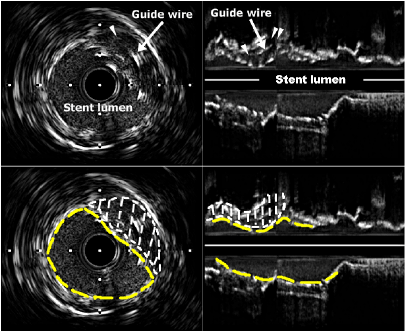
| Figure 3: IVUS images (left panel, cross-section; right panel, longitudinal section) obtained after kissing predilatation of stent and cushion balloon (refer to Fig. 1C); note the under-expanded stent and guide wire (arrow) within the expanded lumen (single arrowhead and area surround by dot, both panels), resulting from cushion balloon inflation, and the patent LCX orifice (double arrowheads, right panel). IVUS, intravascular ultrasound; LCX, left circumflex artery; Yellow dash line, stent struts. |