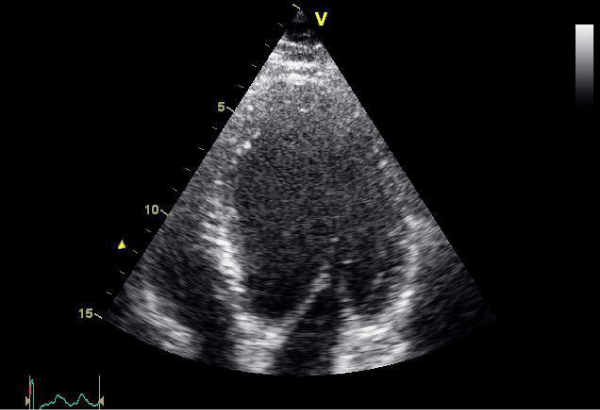
Figure 2:
Echocardiogram (apical view) showing dilated LV.