|
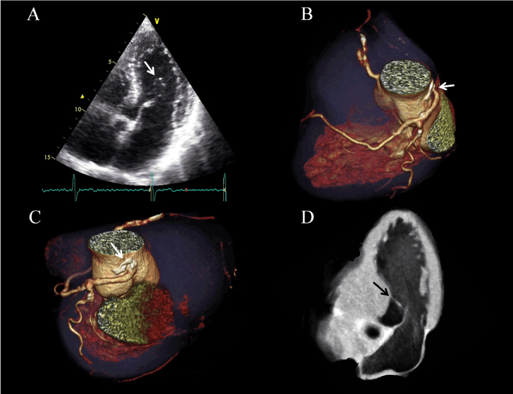
| Figure 1: (A) Two-dimensional echocardiography: apical four-chamber view showing accessory mitral valve tissue (white arrow) connected to the anterior leaflet, floating in the ventricular cavity in diastole. (B-C) Cardiac computed tomography (cCT)-3D reconstruction- presenting the anomalous origin of the LCA (white arrow) and the huge atherosclerotic plaque extending from the aorta in the LM (white arrow). (D) 3D reconstruction from cCT: anatomical section showing the accessory mitral valve tissue in diastole (black arrow). |