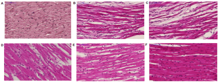
A. Compact structure without any inflammatory infiltrations.
B. Prominent features of cardiomyopathy were seen as cardiomyocytes thinning, stretching, disperse inflammatory cells and interstitial fibrosis.
C. Progressive cardiomyopathy with fibrosis and cardiomyocytes thinning and stretching, inflammatory cells were absent.
D. Cardiomyopathic signs with focal fibrosis and disperse inflammatory cells.
E. Progressive dilation of cardiomyocytes, without progressive fibrosis and inflammation.
F. The signs of cardiomyopathy (i.e. focal fibrosis and cardiomyocytes thinning and stretching) were replaced by cardiomyocytes hypertrophy with slight inflammatory infiltration. Similar changes were visible in group receiving M-2 from 11th to 35th day after MI.