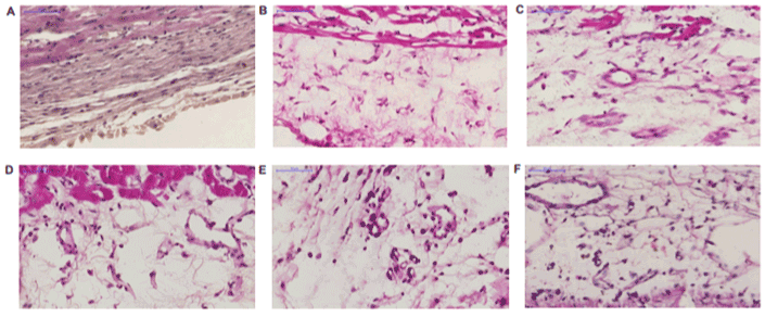
A. Left ventricular myocardium covered by fibrous tissue with single thin-walled vessels.
B. Loose fibrous tissue contains some narrow young vessels, large stimulated fibroblasts and few inflammatory cells. Similar vessels were seen after DMSO administration at 35th day after MI.
C. Thick-walled young capillary vessels were seen between fibroblasts inside young loose connective tissue.
D. Thick-walled young capillary vessels containing endothelial cells with abundant cytoplasm were characteristic inside loose epicardial tissue in this group.
E. More differentiated capillary vessels with scanty cytoplasm and young capillaries with concomitant inflammatory and mast cells.
F. Large young vessels (probably sinusoids) and numerous differentiated small capillaries and maturated fibroblasts. Few inflammatory cells.