|
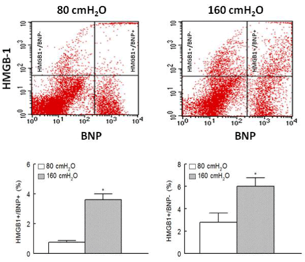
| Figure 1: Panels show representative dot matrices for cardiac cell preparations from ischemic-reperfused hearts, subjected to either 80 or 160 cm H2O, which were positive for HMGB1, with or without reactivity for the brain natriuretic peptide (BNP; a marker of cardiomyocytes). On the other hand, bar graph shows means ± SEM (%) of HMGB1+/BNP+ and HMGB1+/BNP- cells (n=5 hearts/group). * p<0.05 compared to the other group. |