|
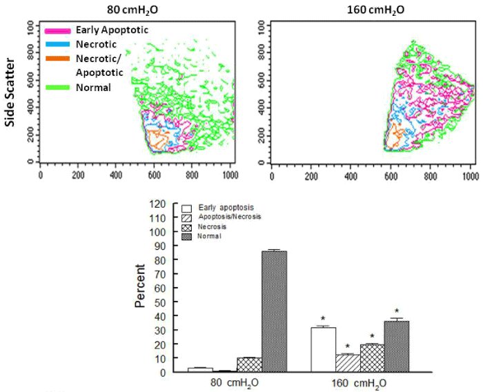
| Figure 4: Panels show representative scatter plots for necrotic, necrotic/apoptotic and early apoptotic cells in cardiac cell preparations from ischemic-reperfused hearts subjected to 80 or 160 cm H2O. Bar graphs show means ± SEM of percent of damaged/dead or normal cells for each group.* p<0.05 compared to the 80 cm H2O group. |