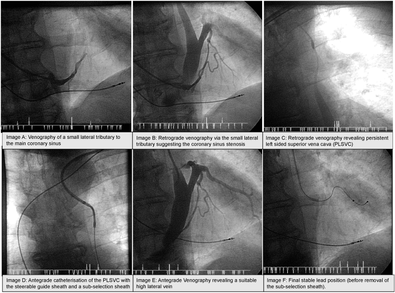
Image A: Venography of a small lateral tributary to the main coronary sinus
Image B: Retrograde venography via the small lateral tributary suggesting the coronary sinus stenosis
Image C: Retrograde venography revealing Persistent Left Sided Superior Vena Cava (PLSVC)
Image D: Antegrade catheterisation of the PLSVC with the steerable guide sheath and a sub-selection sheath
Image E: Antegrade venography revealing a suitable high lateral vein
Image F: Final stable lead position (before removal of the sub-selection sheath).