 |
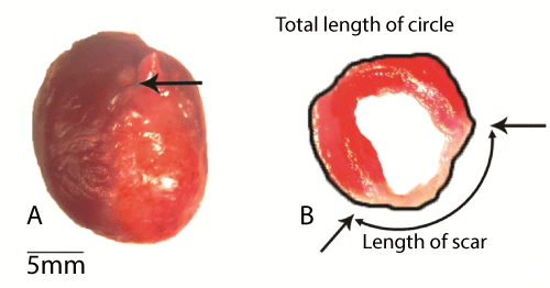
| Figure 1: Images of the LV on the 21st day after LAD occlusion. Panel A: Arrow showing point of ligature. MI hearts revealed irreversible loss of myocardial tissue, and scar formation completely replaced myocardium within the LAD supply region. Panel B: Transverse slice of LV stained with triphenyltetrazolium chloride showing transmural infarction with pronounced wall thinning and aneurysm formation. Percentage of infarcted myocardium calculated as ratio of scar length to total circumference. LAD: Left anterior descending coronary artery; LV: Left ventricle; MI: Myocardial infarction. |