 |
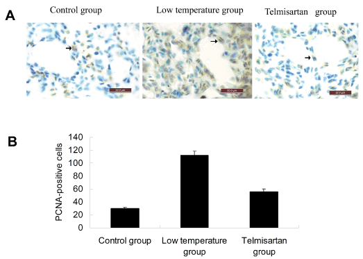
| Figure 4: Effect of telmisartan on PCNA-positive cells in lung tissue. (A) Immunohistochemistry analysis: The PCNA-positive cells were observed under oil lens of microscope. (B) Histograms represent the number of PCNA-positive cells per visual field. The Ang II-positive cells of 20 fields of tubular area at high magnification were counted and averaged. Data are presented as the mean ± SEM (n=8 in each group).**P<0.01 compared with Control group and Telmisartan group(one-way ANOVA). |