|
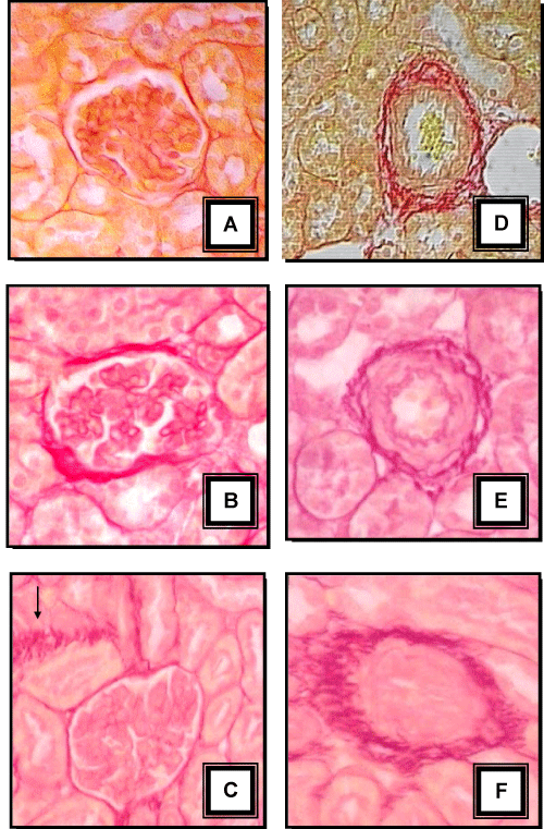
| Figure 2: Representative photomicrographs of the renal cortex from animals from the group control, L-NAME and SHR at 15 days of age. Tissues shown in A, B, C, D, E and F were stained with picrosirius red. There was greater collagen accumulation in the glomeruli of L-NAME (B) and SHR (C) at 15 days of age than control group (A). Excess collagen (arrow) can be seen in the tubular interstitium of SHR neonates (C) and the periadventitia of cortical vessels in L-NAME (D) and SHR (E) animals. Magnification, ×400. |