 |
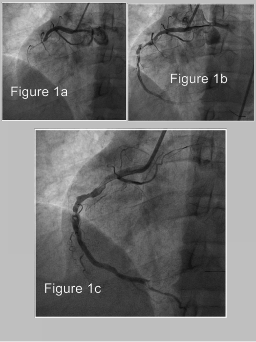
| Figure 1: A) Left anterior oblique view of the right coronary artery (RCA) revealing 100% stenosis of the proximal artery. B) Left anterior oblique view of the RCA revealing the TIMI grade 1 flow restored just after crossing the proximal lesion. High thrombus burden of vessel spreading from proximal segment to the acute margine level and the slight tortuosity of the artery are clearly visible. C) TIMI grade 3 flow restored after the suction of proximal thrombus in RCA. Note: The distal posterolateral branch was occluded because of distal thrombus embolization. |