 |
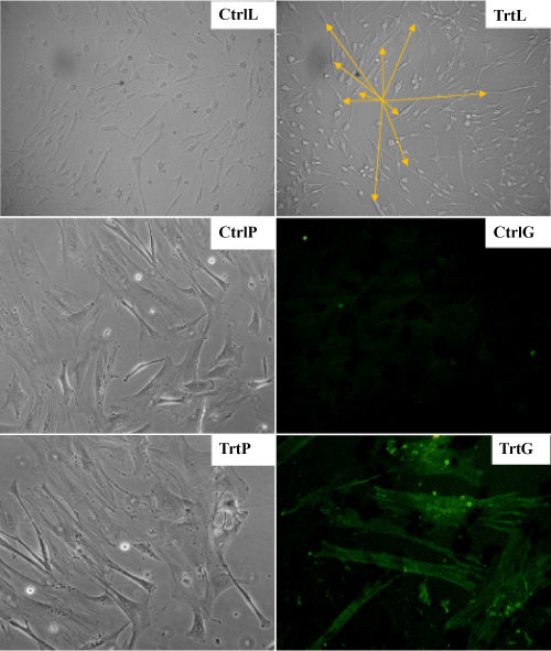
| Figure 1: aFRLM-enhanced cardiogenic differentiation of cultured MSCs. The majority of the vehicle-treated MSCs (14 days) under low power filed (CtrlL) and high power filed (CtrlP) phase contrast microscope remained flat, irregular, asymmetry and low refractive with few MHC positively (GFP) stained cells (CtrlG). By contrast, approximately 30% of the cultured MSCs in aFRLM-treated wells (14 days) under low power filed (TrtL) and high power filed (TrtP) became relatively elongated and high refractive myocyte-like phenotype (yellow arrow heads) with approximately 30% of these myocytelike cells were positively (GFP) stained with MHC specific antibodies (TrtG). |