|
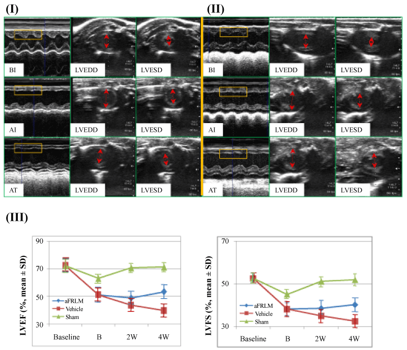
| Figure 2: Echocardiography assessment of cardiac function. Representative M-mode and end diastolic/end systolic short axis cross sectional images of the left ventricle at the level of the papillary muscle of aFRLM-treated (I) and vehicle-treated (II) rat MI hearts. LVEDD: left ventricular end diastolic diameter; LVESD: left ventricular end systolic diameter; BI: normal rat echocardiography before MI induction; AI: images taken three days post infarction, but prior to any treatment; AT: images taken 4 weeks post either aFRLM or vehicle treatment respectively. Before the LAD ligation surgery, echocardiograms of animals in both groups showed a normal motion of structure throughout several cycles in M-mode measurements of a short axis view (I:BI & II:BI) (yellow rectangular enclosed) and normal LVEDD and LVESD (upper panels in I and II). Three days post ligation of LAD (I:AI & II:AI), anterior akinesis (yellow rectangular enclosed) and marked left ventricle dilation (red arrow heads) were observed in both groups of animals (middle panels in I & II). Four weeks of aFRLM treatment progressively improved the motion of left ventricle anterior walls (yellow rectangular) and reduced diastolic/systolic diameter (red arrow heads) (lower panel in I). By contrast, the vehicle-treated control hearts (II) remained akinesis (yellow rectangular) and progressively increased in diastolic/systolic diameter (red arrow heads) (lower panel in II). III, LVEF and LVFS measurements demonstrated the improved heart function with time by aFRLM treatment. Vehicle: vehicle-treated group; aFRLM: aFRLM-treated group; sham: open chest surgery without LAD ligation group; baseline: before ligation; B: 3 days post ligation; 2W: 2 weeks after aFRLM or vehicle treatment; 4W: 4 weeks after aFRLM or vehicle treatment. The graphs showed the restoration of the cardiac function in aFRLM-treated, but not vehicle-treated hearts. |