|
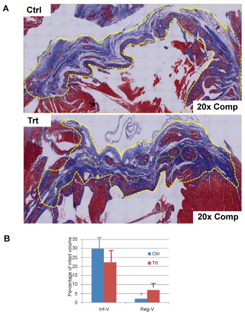
| Figure 3: Morphometrical assessment of therapeutic effect of aFRLM on MI. A: The representative figures with the whole cross-field of infarction stained by Masson’s trichrome method were composed by 130 (Trt) and 160 (Ctrl) consecutive microscopic photos (20X). The vehicle-treated MI heart (Ctrl) showed a bluestained large and thinned infarct area (yellow dashed line surrounded). The fibrous scar of the infarct was stained blue. By contrast, aFRLM-treated MI heart showed a smaller and less thinned infarct area (yellow dashed line surrounded). More interestingly, many red-stained myocyte-like cell clusters (green circles) replaced the blue-stained fibrous scar and reduced the infarct volume in aFRLM-treated MI heart. B: Inf-V, the infarct volumes in both aFRLM- (Trt) and vehicle- (Ctrl) treated hearts. Reg-V, the regenerating myocyte volumes in hearts of both groups. Note, some newly formed cardiac myocyte clusters (A: green circles) replaced about one fourth (6.9 ± 2.3%) of the original necrosed cardiac tissues reducing the average infarct volume to 22.3 ± 7.0% of the left ventricle volume in aFRLM-treated group. By contrast, the average infarct volume occupied about 29.8 ± 7.3% with less than 2.1 ± 1.1% regenerating myocyte-like cells observed in vehicle-treated hearts (P<0.01). |