|
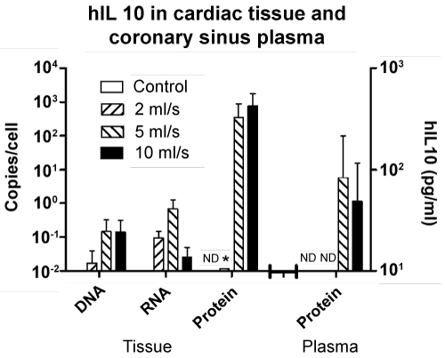
| Figure 3: hIL-10 gene delivery and information decoding process in non-infarcted cardiac tissue. After infarct induction, 50 ml of saline solution containing hIL-10 plasmid was injected retrovenously at 2, 5 or 10 ml/s into the coronary sinus. After 72 hours, pigs were sacrificed and tissue samples throughout the heart were collected for molecular analyses. Quantitative PCR, RT-PCR and ELISA of exogenous hIL-10 gene and transcript in samples of non-infarcted cardiac tissue were performed, and results were expressed as hIL-10 DNA, RNA and protein copy number per cell. Protein expression in plasma was expressed as concentration in pg/ml. ND: undetermined; * it was not possible to evaluate. |