 |
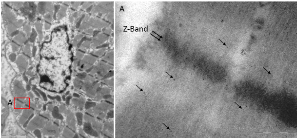
| Figure 5: Gold nanoparticle distribution in hepatocytes. A 50 ml volume of citric buffer solution containing gold nanoparticles with diameters of 4 nm and 15 nm (1012 nanoparticles of each size per ml) was injected into the heart at 5 ml/s. The image at left shows a cardiomyocyte with its nucleus. Box A corresponding to the squared area is enlarged (image at right). Only 4 nm diameter particles are present within the cell, especially surrounding the Z-band area. The 4 nm particles are widely distributed within the cytoplasm, while the larger nanoparticles appear to be less abundant. |