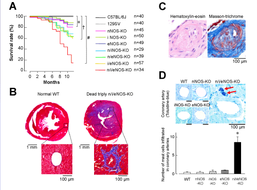
(A) Survival rate (n=29-57). The red line represents markedly reduced survival in the triply n/i/eNOSs-/- mice. *, †, and #: P<0.05 between wild-type (WT) C57BL/6J vs. singly, doubly, and triply NOS-KO, respectively. (B) Acute MI and coronary arteriosclerotic lesion formation in a triply n/i/eNOSs-/- mouse that died at 8 months of age (Masson-trichrome staining). Blue in the heart cross-section of the dead triply n/i/eNOSs-/- mouse indicates antero-septal acute MI. Adjacent coronary artery shows marked luminal narrowing, wall thickening, and perivascular fibrosis (blue). (C) Arteriosclerotic lesion formation in serial sections of the infarct-related coronary artery. (D) Mast cell infiltration in the coronary artery adventitia (toluidine-blue staining) (n=10-33). Red arrows indicate mast cells. *P<0.05 vs. WT [62].