|
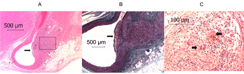
| Figure 3: Coronary artery treated with 600 μg collagenase at 30 d post injection. There was a prominent intimal thickening on the ballooned treated site of the coronary artery (indicated by black arrows in panel A and B) and a marked inflammatory response adjacent to coronary artery. Box in panel A indicates eosinophil’s, macrophages and giant cell infiltration, which is magnified in panel C. Black arrow in panel C indicate the multi nucleated giant cell infiltration. |