 |
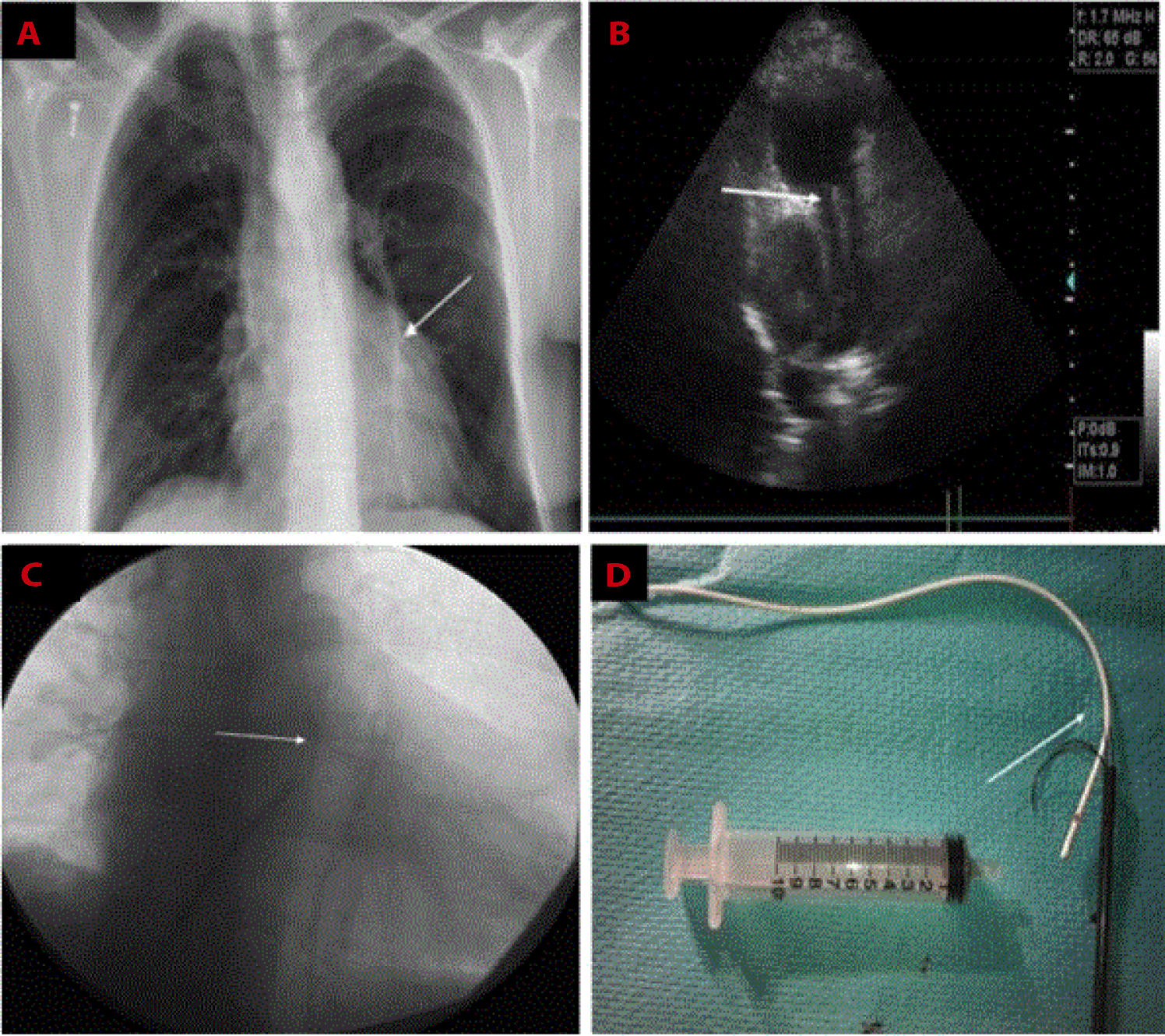
| Figure 1: A- Initial chest film showing migrated catheter fragment into the left pulmonary artery (white arrow). B- Trans-thoracic echocardiography visualising catheter fragment in the left pulmonary artery (white arrow). Right pulmonary artery is free. CThe tip of the fragment in the left pulmonary artery is captured with The Needle's Eye Snare® (white arrow). D-The vascular sheath and the 23 cm snared catheter fragment were withdrawn as a unit out through the skin (white arrow). |