 |
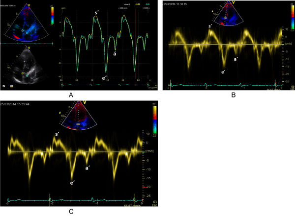
| Figure 1: a-c)The systolic (s´), early diastolic (e´) and late diastolic (a´) velocities measured at a septal and at a lateral site of the aortic annulus (AA) by using quantitative two-dimensional color Doppler tissue Imaging (TVI) (a) and by using pulsed-wave Doppler tissue imaging at the septal site of AA (b) and from a lateral site of the AA (c) from the apical five-chamber view in a 28-year-old healthy woman. |