|
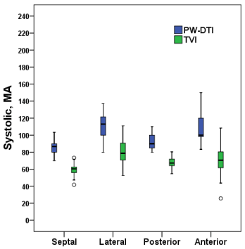
| Figure 2b: Box plots showing systolic velocities measured with PW-DTI and TVI at four different sites at the mitral annulus. Boxplot is defined by median, quartiles (box), min-max (whiskers) and circle markers if outliers are present. (MA=mitral annulus, PW-DTI=pulsed wave tissue Doppler imaging, TVI=colour coded tissue Doppler imaging, CI=confidence interval) (n=24). |