 |
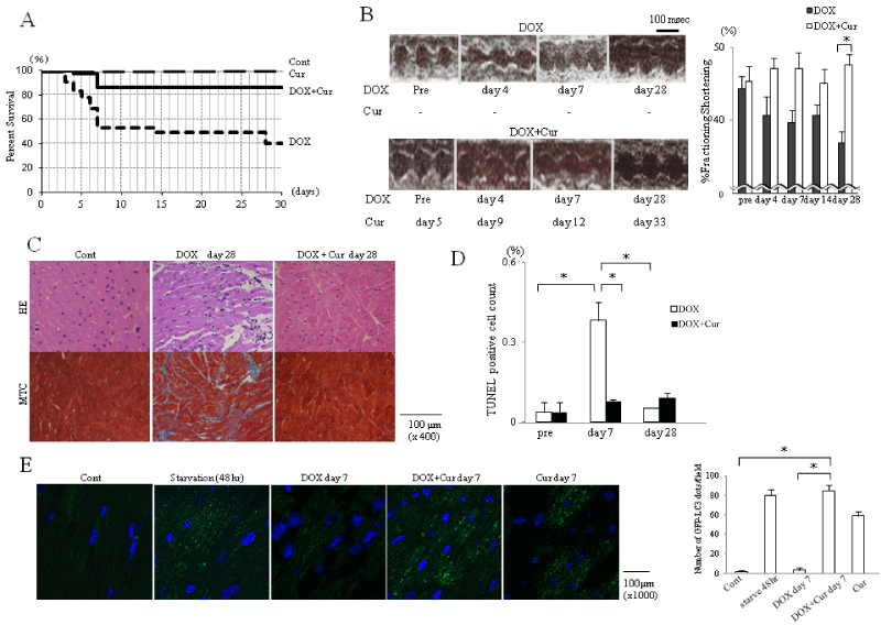
| Figure 1: Curcumin protects C57Bl /6 mice heart from doxorubicin induced heart failure. (A) Cur-treated mice group with DOX showed drastic survival benefit. Cont; n=10, survival rate 100%, Cur; n=10, 100%, DOX+Cur; n=37, 87%, DOX; n=77, 40% Survival rate of DOX group on day 7 was 52%. (B) Left, Typical time course of M-mode echocardiography of both groups measured LV function at 4, 7, 28 days after DOX injection. Cur was started 5 days prior to DOX. Right, Sequential change of % FS in each group. Each bar represents mean ± SEM. *p<0.01, (n=6 per group). (C) Microscopic pathology of both group mice heart. Hematoxylin-Eosin (HE) staining and Masson trichrome (MTC) staining of mouse myocardium illustrate cardiac fibrosis, disarray as well as atrophic change of cardiomyocyte in DOX treated mice heart on 28 days. Cur suppresses these changes in DOX-treated mice myocardium. (n=6 per group). (D) In quantification of the TUNEL positive cells count, each bar represents mean ± SEM of 3 fields. *p<0.01 (n=6 per group). (E) Microscopic photograph of cardiomyocyte of GFP-LC3 transgenic mice illustrating green GFP-LC3 dots. Nuclei are stained blue with DAPI. In quantification of the GFP-LC3 dots per fields, each bar represents mean ± SEM of six fields. *p<0.01 Cont: Control; DOX: Doxorubicin, Cur: Curcumin. |