 |
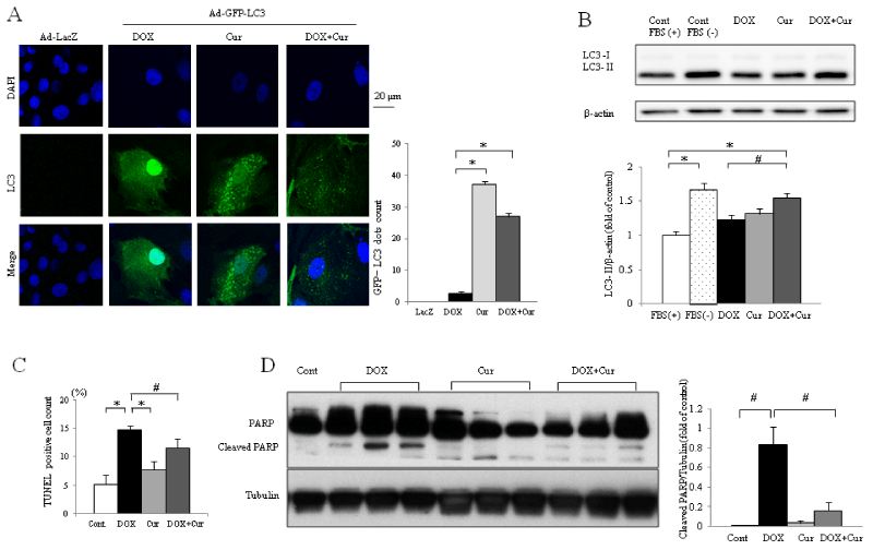
| Figure 2: Curcumin induces autophagy and reduces apoptosis in neonatal rat cardiomyocyte after Doxorubicin treatment. (A) Confocal photomicrograph of cardiomyocyte infected with GFP-LC3-expressing adenovirus. In quantitation of GFP-LC3 puncta formation per cell, LC3 puncta were counted in representative section of the sample. Each bar represents mean ± SEM of six independent experiments. *p<0.01 LacZ ; control Ad-LacZ gene transfer. (B) Western blot analysis of LC3. Data were normalized with β-actin which serves as a loading control. Each bar represents mean ± SEM of six independent experiments. *p<0.01, #p<0.05 (n=4 per group). (C) Data shown are averages of TUNEL staining of cardiomyocyte in eight fields per condition of five time experiments. Each bar represents mean ± SEM of six independent experiments. *p<0.01, #p<0.05. (D) Representative western blot analysis to detect cleaved PARP in neonatal rat cardiomyocyte. Data were normalized with Tubulin which serves as a loading control. Each bar represents mean ± SEM of four independent experiments. #p<0.05 Cont: Control; DOX: Doxorubicin, Cur: Curcumin. |