|
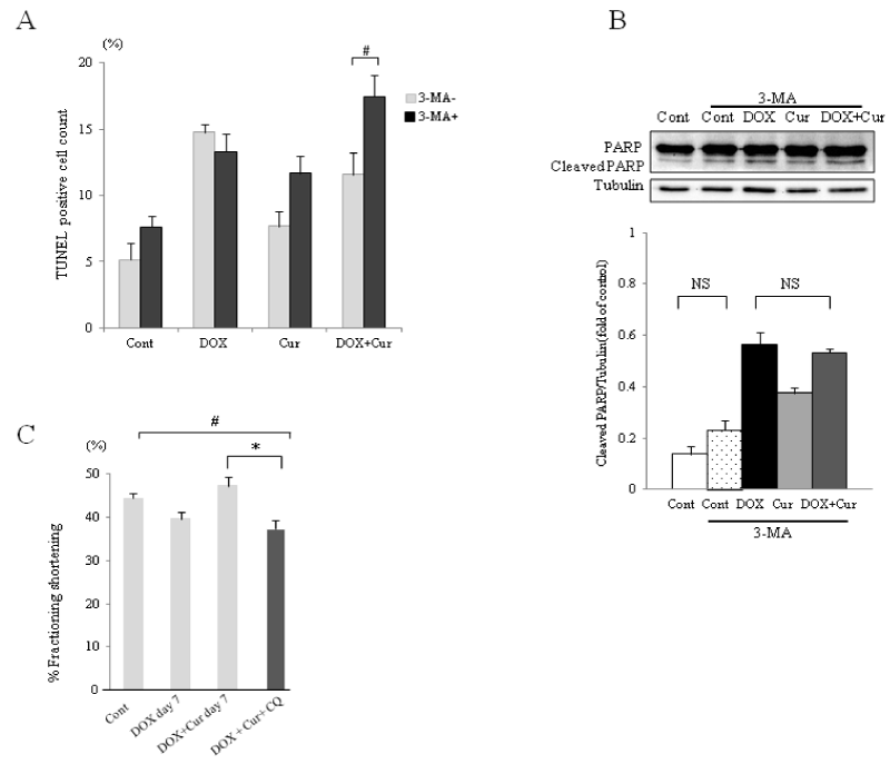
| Figure 3: Inhibition of autophagy prevents curcumin from protecting cardiomyocyte. (A) Quantitation of TUNEL positive cell nuclei counts of each group. Data shown are averages of eight fields per condition of eight times experiments. Data are mean ± SEM. #p<0.05. (B) Representative western blot analysis to detect cleaved PARP in neonatal rat cardiomyocyte treated with 3MA. Data were normalized with Tubulin which serves as a loading control. Each bar represents mean ± SEM of four independent experiments. (n=4 per group). (C) %FS measured by M-mode echocardiography at 7 days after DOX injection of DOX+Cur-treated mice with chloroquine. Cur was started 5 days prior to DOX. Data are mean ± SEM. (n=6 per group) *p<0.01 #p<0.05. Cont: Control; DOX: Doxorubicin, Cur: Curcumin; NS: Not Significant. |