 |
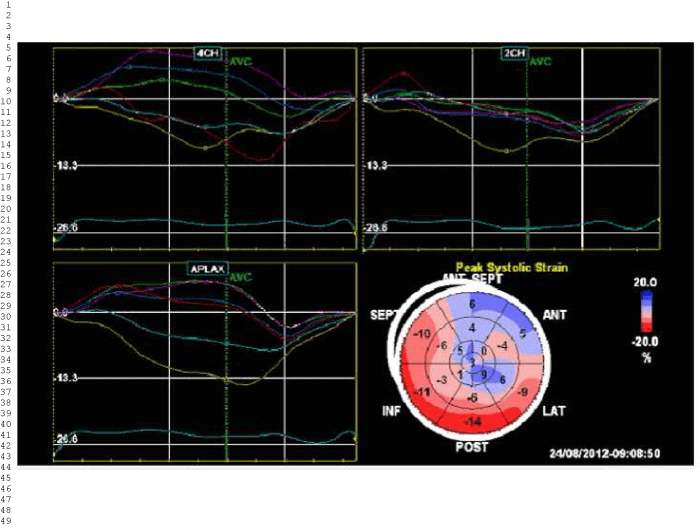
| Figure 6: Strain analysis of this bullseye view shows hypokinesis in the anterior and apical regions of the left ventricle (strain 0 to 9%). Basal, septal, inferior and posterior regions appear to maintain kinesis at low normal range in figures 4 and 5. |