|
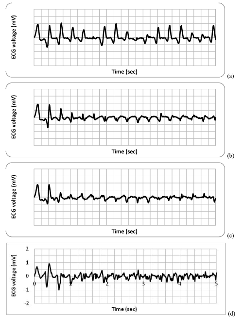
| Figure 7: Simulated ECG tracings for VT/VF in models of increasing complexity in the endocardial to epicardial dimension. (a) Purknje fibers on and myocardial gradients off. (b) Endo to epicardial gradient of ERP is added. (c) Endo to epicardial gradient in conduction velocity in the abnormal region is also added with endocardial velocity being faster. (d) Lumped Purkinje fibers added to create the standard model. |