|
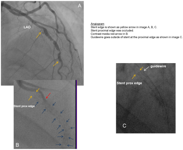
| Figure 2: A) An angiogram was performed and the two yellow arrows demonstrate the stent edges with in stent restenosis being evident. B) Closer analyses reveal that the proximal stent edge was occluded (yellow arrow) as contrast seems to be outside the stent (red arrow). The blue arrows outline the stent location throughout the vessel. C) In this close-up of the vessel, it is demonstrated that the guide-wire travels outside the proximal edge of the stent. |