|
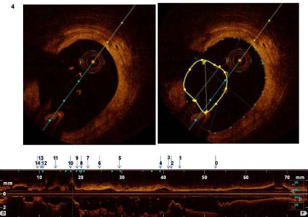
| Figure 3: The top panel demonstrates the cross-section of the vessel and the bottom panel is a longitudinal representation of the entire length of the vessel being scanned. The arrows demonstrate the location in terms of the vessel. In this case, position #4 in the vessel reveals a positively remodelled vessel with stent malapposition. (The yellow circle encompasses the circumference of the stent which is much smaller that the circumference of the entire vessel). |