 |
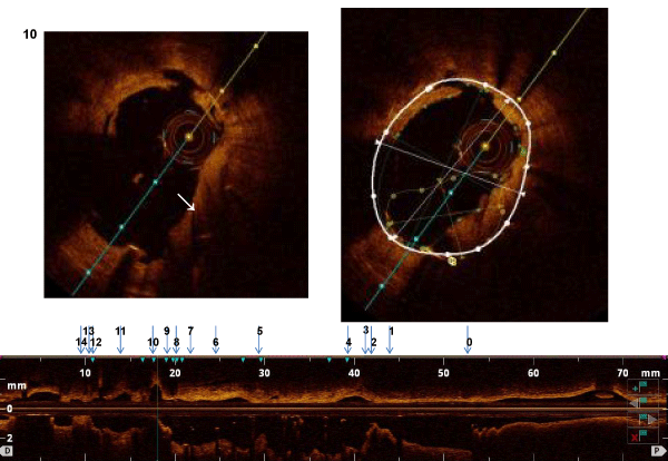
| Figure 5: The top panel demonstrates the cross-section of the vessel and the bottom panel is a longitudinal representation of the entire length of the vessel being scanned. The arrows demonstrate location in terms of the vessel. In this case, position #10 in the vessel reveals a positively remodelled vessel with stent malapposition. (The yellow circle encompasses the circumference of the stent which is much smaller that the circumference of the entire vessel (white circle)). In stent restenosis is also evident (white arrow). |