|
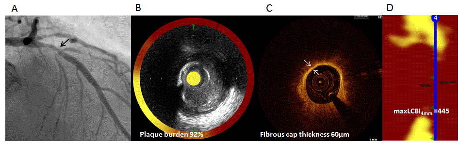
| Figure 3: The Power of Multimodality Imaging. A) Angiogram of 59 year old patient presenting with unstable angina. Lesion (arrow) in LAD (left anterior descending artery) identified; B) Intravascular ultrasound (IVUS) of same segment demonstrates total plaque burden; C) Optical coherence tomography (OCT) reveals thin cap fibroatheroma (see arrows); D) Near infrared spectroscopy (NIRS) provides information regarding lipid content. |