|
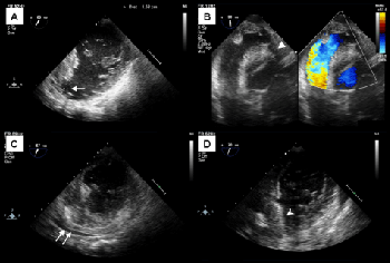
| Figure 1: TEE: (a) modified mid-esophageal mitral commissural view showed the VSD concomitant with an apical aneurysm (arrow). (b) Deep-transgastric long-axis view showed an apical VSD (arrowhead) with left-to-right shunting. (c) Modified midesophageal mitral commissural view demonstrated pericardial effusion (double arrow) after LV wall perforation. (d) Modified mid-esophageal mitral commissural view showed that pericardiac effusion was relieved after pericardiostomy and surgical repair without Amplatzer occluder displacement (hollow arrowhead). |