 |
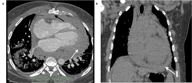
| Figure 1: Axial and coronal chest CT images respectively. The axial image demonstrates the diffuse, thickened pericardium and associated effusion (white arrow), despite the patient’s previous sternotomy (black arrow). This finding is also readily identified on the coronal image. A visible pericardial mass was not identified on either section. |