 |
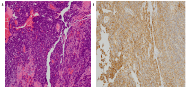
| Figure 2: Histological specimens obtained from the pericardium (10X). The lesion shows exuberant proliferation of cells with high nuclear to cytoplasmic ratio, numerous mitoses, and necrosis with focal papillary architecture and anastomosing vascular channels in the first image. The second image demonstrates positive immunohistochemical stain for CD31, a marker found on endothelial cells and associated with vascular tumors such as angiosarcomas |